|

Fig.
1

Fig.
2

Fig. 3 |
One of the most popular
techniques for clinical investigation of the retina is retinal
imaging. It is usually performed by specialized instrument -
fundus camera.
The conventional fundus cameras work with an input pupil of
about 2mm to reduce the negative influence of eye aberrations.
However, using a larger input pupil and introducing adaptive
correction of aberrations, it is possible to achieve the
resolution of the cell level range. The first results of such
adaptive correction were presented by
Prof. D.Williams and co-workers.
The information content of fundus images can be further enhanced
by spectral-resolved photography called
multi- or hyper-spectral
imaging.
With a high-resolution
spectral imager, a broad range of eye diseases that represent
the leading causes of visual impairment and/or blindness can be
studied in-vivo.
For example, in diabetes, a microvascular disease that causes
retinopathy, the proposed device may enable the investigation of
detail changes in individual capillary beds that occur early in
the disease within the inner retina. At spatial resolutions that
will provide unprecedented detail of capillary beds
(5-10 um),
one will be able to make measurements of oxyhemoglobin
saturation and detect ischemic regions. This resolution is
nearly ten times
better than can be achieved by existing fundus cameras which can
not compensate for the unique aberrations in each patient's eye.
Together with
Kestrel Corporation
(Albuquerque, US) our group was involved in the development of
the SRSFI
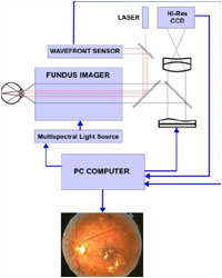
(Super Resolution Spectral Fundus Imager) system. The work was
supported by NATO SfP Grant #974292. The basic layout of the
system is shown in
Fig. 1.
The system includes a
Shack-Hartmann
wavefront sensor, a
bimorph adaptive mirror with 18 electrodes, a hi-resolution
digital camera (3000x2000 pixels, 12 bit), and a multispectral
light source with 8 bands (from 80 to 8nm). A more detailed
description of the system can be found in US Patents: US patent
#US
6331059 B1 and
#US
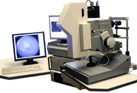
2002097377 A1. A snapshot of the system is shown
in Fig. 2.
The system works with an
input pupil of 5 mm
in diameter and the
typical residual error of correction is
0.1 um
(RMS). The spatial resolution of the system is 6mcm on the
retina (limited by the CCD sensor). The angular field of view is
15x20 deg.
A more detailed technical specification of the device can be
found [
here
].
Two SRSFI systems have been built in the framework of the NATO
SfP Grant #974292. The systems are almost identical. The first
has been installed in Albuquerque (Kestrel Corp.) and the second
in Moscow (Medical
Physics Department, Faculty of physics, Moscow
State University).
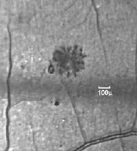
An example of a retinal
image taken by the SRSFI system is shown in Figure 3 (only a
portion of the larger image is presented).
Presently, the second
generation of the SRSFI instruments (SRSFI-II) is being
developed (Fig. 4).
The new instrument has a more advanced wavefront sensor and
adaptive optics control with a 77Hz loop rate. The stroke of the
bimorph mirror is increased up to 36 microns. A large-format 16
MPix data camera with a high QE sensor from Kodak has been
integrated into the system. This makes it possible to increase
the spatial resolution
by 30% without
limiting the angular FOV. A more detailed information about the
SRSFI-II can be found
[
here
].
An SRSFI-II system is
installed in the
Research Institute of Eye Diseases,
Moscow, Russia, where limited clinical trials of utility of the
SRSFI for diagnostics and management of several retinal diseases
are being carried out.
The main focus of the
current research is now on methods of image analysis and
restoration under
angular anisoplanatism
conditions common for the human eye. Starting from our earlier
work we developed several
methods for extending the FOV in human eye adaptive imaging. The
scanning reference source employed in the SRSFI-II extends the
FOV by 30-50%. This approach is quite similar to the multiple
beacons AO. At the same time, wide-angle
noniterative blind deconvolution helps to further
extend the system FOV. As a result, the compensated FOV of the
SRSFI-II is about 15
deg.
|
|
PUBLICATIONS:
A. Larichev, P. Ivanov, I. Irochnikov, V. Shmalhauzen, L.J.Otten,
Adaptive system for eye-fundus imaging,
Quantum Electronics, 32, №10 (2002)

A. Larichev, P. Ivanov, I.
Irochnikov, S.C. Nemeth, A. Edwards, P. Soliz, High Speed
Measurement of Human Eye Aberrations with Shack-Hartman Sensor.
[ARVO Abstract]
Invest Ophthalmol Vis Sci., 42 (2001) 897

A.V.Larichev, P.V.Ivanov,
I.G.Irochnikov, V.I.Shmal'gauzen, Measurement of eye aberrations
in a speckle field, Quantum Electronics, 31 (2001) 1108

P. Fournier, G. R. G.
Erry, L. J. Otten, A. Larichev, N. Irochnikov, A Next Generation
High Resolution Adaptive Optics Fundus Imager. Article 
P. Fournier, G. R. G.
Erry, L. J. Otten, A. Larichev, N. Irochnikov, A Next Generation
High Resolution Adaptive Optics Fundus Imager. Presentation

A.V. Larichev, J.J. Otten,
N.G. Irochnikov, P. Soliz, G.R.G. Erry, V.Y. Panchenko,
SuperRez-II adaptive multispectral fundus imager, SPIE, 6138-38
V.1

|